Generic selectors
Exact matches only
Search in title
Search in content
Post Type Selectors
Filter by Categories
Acknowledgements
Authors’ reply
Book Review
Book Reviews
Classics In Indian Medicine
Clinical Case Report
Clinical Case Reports
Clinical Research Methods
Clinico-pathological Conference
Clinicopathological Conference
Conferences
Correspondence
Corrigendum
Editorial
Eminent Indians in Medicine
Errata
Erratum
Everyday Practice
Film Review
History of Medicine
HOW TO DO IT
Images In Medicine
Indian Medical Institutions
Letter from Bristol
Letter from Chennai
Letter From Ganiyari
Letter from Glasgow
Letter from London
Letter from Mangalore
Letter From Mumbai
Letter From Nepal
Masala
Medical Education
Medical Ethics
Medicine and Society
News From Here And There
Notice of Retraction
Notices
Obituaries
Obituary
Original Article
Original Articles
Review Article
Selected Summaries
Selected Summary
Short Report
Short Reports
Speaking for Myself
Speaking for Ourselve
Speaking for Ourselves
Students@nmji
View/Download PDF

Translate this page into:

Images In Medicine
2017:30:4;241-241
doi: 10.4103/0970-258X.218685
PMID: 29162763

Incidental cytodiagnosis of microfilaria from subcutaneous nodule

T Santosh1 , Hemlata Panwar2 , Amit Bugalia2 , Vandita Yogendra Singh2 , Nighat Hussain2
1 Department of Pathology, Seth G.S. Medical College and K.E.M. Hospital Parel, Mumbai, Maharashtra, India
2 Department of Pathology and Laboratory Medicine, All India Institute of Medical Sciences, Raipur, Chhattisgarh, India

Corresponding Author:
Hemlata Panwar
Department of Pathology and Laboratory Medicine, All India Institute of Medical Sciences, Raipur, Chhattisgarh
India
hemlatasongra@gmail.com
How to cite this article:
Santosh T, Panwar H, Bugalia A, Singh VY, Hussain N. Incidental cytodiagnosis of microfilaria from subcutaneous nodule. Natl Med J India 2017;30:241
Copyright: (C)2017 The National Medical Journal of India

A 35-year-old woman presented with a swelling of the right forearm to the surgery outpatient department at the All India Institute of Medical Sciences, Raipur. On examination, she had a subcutaneous swelling in the right forearm, painful and diffuse measuring 3× 3 cm for 6 months [Figure - 1]A. A fine-needle aspiration was done and the material was sent for cytological examination with a clinical impression of fibro-lipoma. Blood mixed fluid was aspirated and routine Giemsa and Pap stains were done. Cytological examination revealed epithelioid granulomas with lymphocytes and microfilariae in a haemorrhagic background [Figure - 1]B, [Figure - 1]C, [Figure - 1]D. The microfilaria was sheathed and the column of nuclei in them with the tail tip being free [Figure - 1]E, [Figure - 1]F. A cytological diagnosis of granulomatous inflammation due to microfilaria morphologically consistent with Wuchereria bancrofti was made. She was treated with diethylcarbamazine (DEC) for 3 weeks. On follow-up, the swelling subsided.

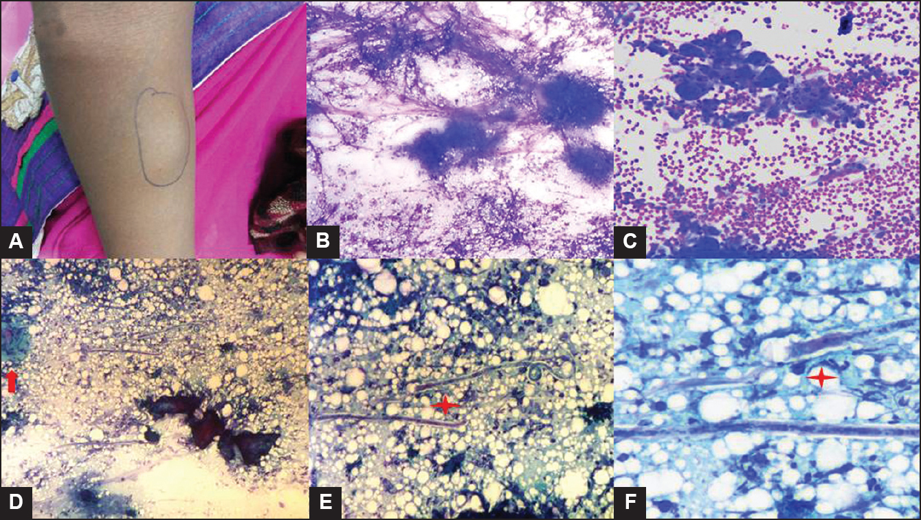
Figure 1: Swelling in the right forearm of the patient Figs1B, C, D, E, F. Cytosmears showing microfilariae (star) in a granulomatous inflammatory (arrow) background comprised foreign body-type giant cells and histiocytes (Giemsa × 400; PAP, × 400)

Even in the absence of clinical indications and eosinophilia in peripheral smears, microfilaria can be detected in subcutaneous nodules. Hence, careful screening for microfilariae is important in aspiration cytology smears.


Fulltext Views
770

PDF downloads
591
Show Sections