Translate this page into:
Pigmented squamous cell carcinoma of the conjunctiva
Corresponding Author:
K R Sheera
Chaithanya Eye Hospital and Research Institute, Thiruvananthapuram, Kerala
India
sheeraarun@gmail.com
| How to cite this article: Sheera K R, Kurian A. Pigmented squamous cell carcinoma of the conjunctiva. Natl Med J India 2019;32:316 |
A 79-year-old male, not a diabetic or hypertensive, presented to the outpatient department with pigmented painless lesions in the temporal quadrant of his right eye for 3 years without visual impairment. The intraocular pressure was normal and the media was clear. The two melanomatous lesions were firm, and nodular with neovascularization [Figure - 1]a. The temporal lesion was freely mobile. The limbal mass with restricted mobility was excised for biopsy. Histopathological evaluation suggested multiple foci of carcinomatous proliferation involving the squamous epithelium lining the limbal nodule with typical keratin pearls [Figure - 1]b. The sub-epithelial tissues showed several melanophores and lymphocytic infiltration with neovascularization, suggestive of moderately differentiated pigmented squamous cell carcinoma of the bulbar conjunctiva, a rare entity. Metastatic work-up was negative. Ultrasound biomicroscopy excluded involvement of the ciliary body and iris [Figure - 2]. The other conjunctival mass was widely excised with 4–6 mm margins using the ‘no-touch’ technique, cryotherapy of the conjunctival margins, alcohol kerato-epitheliectomy and amniotic membrane transplantation with glue under local anaesthesia. The patient remains well at 3 years’ follow-up [Figure - 3].
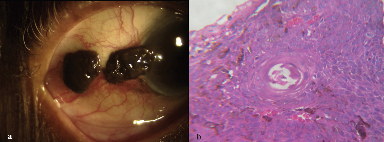 |
| Figure 1: (a) The melanomatous irregular nodular lesions in the temporal conjunctiva of the right eye with increased vascularity; (b) histopathological evaluation of the lesions showing keratin pearls and infiltration of the conjunctival epithelium with moderately differentiated malignant cells and plenty of melanophores (H and E, ×400) |
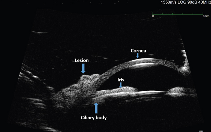 |
| Figure 2: Ultrasound biomicroscopy of the right eye showing the lesion in the limbal region without evidence of invasion of the iris or ciliary body |
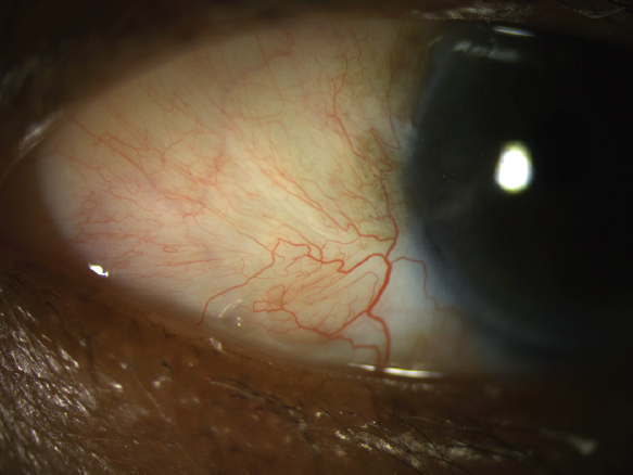 |
| Figure 3: Follow-up image of the right eye at 3 years showing no recurrence |
Conflicts of interest. None declared
Acknowledgement
We thank Vinay S. Pillai and Late M. Balaraman Nair for their contribution to patient care
Fulltext Views
1,137
PDF downloads
357


![[Figure - 1]](#fig_NatlMedJIndia_2019_32_5_316_295964_f1.jpg)
![[Figure - 2]](#fig_NatlMedJIndia_2019_32_5_316_295964_f2.jpg)
![[Figure - 3]](#fig_NatlMedJIndia_2019_32_5_316_295964_f3.jpg)