
Translate this page into:
Cortical versus nuclear lenticular fluid
[To cite: Bhayana AA, Khokhar S. Cortical versus nuclear lenticular fluid. Natl Med J India 2024;37:050. DOI: 10.25259/NMJI_1069_2022]
Crystalline lens is like a dustbin where older lens fibres keep accumulating in the centre on account of continuous influx of fresh fibres from the anterior equator, thereby causing central sclerosis. Lenticular fluid accumulation1 is a form of cataract that behaves differently in the cortex and in the nucleus on account of differential compactness of fibres. We document the same in a 50-year-old woman with diabetes who, in her right eye had amoeboid and milky fluid pocket (anterior subcapsular region) (Figs 1a,b––red circle; Fig. 1c––red arrow), whereas in the left eye, she had compact clear vesicles (nucleus) (Fig. 1d––green circle; Fig. 1e––green arrow).The two have unique implications. It is always the milky superficial fluid that seeps out in the anterior chamber during anterior capsulorhexis opening. Though it opacifies the field of rhexis view, it decompresses the lenticular volume, thereby reducing chances of rhexis runoff/extension. A calculated nick over the same area to begin rhexis can help the surgeon take a chance of Argentinian flag sign in intumescent cataracts. Nuclear fluid pockets, on the other hand, are sources of optical distortions and may warrant surgery in the absence of any gross lenticular opacity or sclerosis.
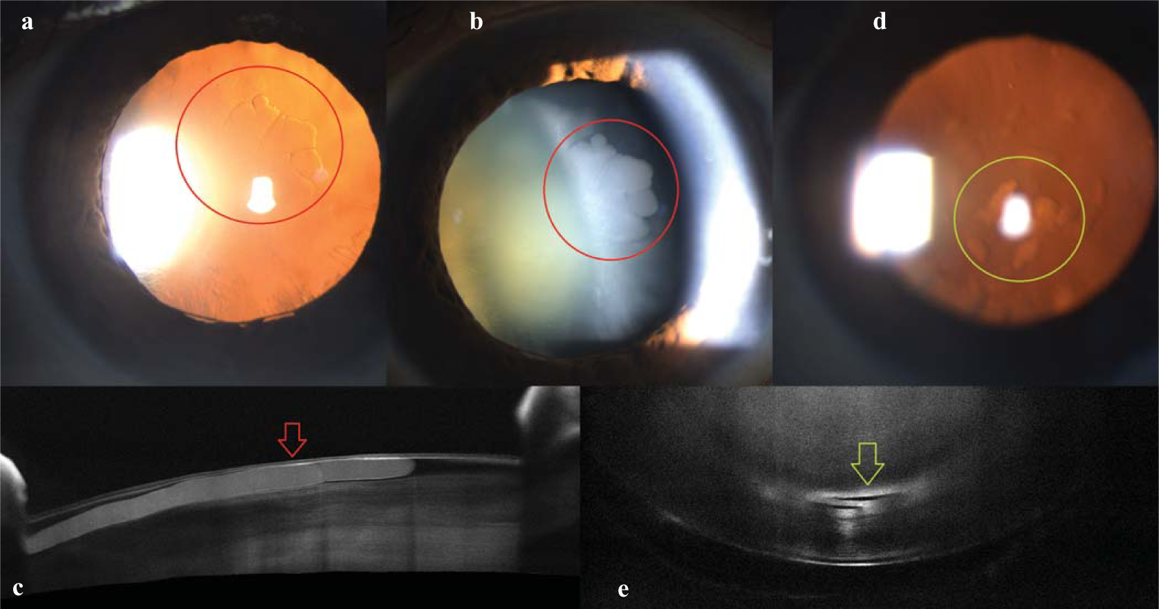
- (a,b) Right eye fluid pocket––red circle; (c) right eye fluid pocket on anterior segment optical coherence tomography––red arrow; (d) left eye fluid pocket––green circle; (e) left eye fluid pocket on anterior segment optical coherence tomography––green arrow
References
- Associations of human crystalline lens retrodots and waterclefts with visual impairment: An observational study. Invest Ophthalmol Vis Sci. 2002;43:2105-9.
- [Google Scholar]


