
Translate this page into:
Detached Schwalbe line in Axenfeld–Rieger syndrome
[To cite: Pillai M, Pabolu C, Chaudhary S. Detached Schwalbe line in Axenfeld–Rieger Syndrome. Natl Med J India 2024;37:230. DOI: 10.25259/NMJI_382_2023]
A 6-year-old boy with Axenfeld–Rieger syndrome presented with complaints of watering in both eyes. His visual acuity was 6/36 in the right eye and 6/9 in the left eye, along with an intraocular pressure of 44 mmHg and 18 mmHg, respectively. Examination revealed megalocornea, a white, cord-like structure resting on the anterior surface of the iris with attached iris strands representing a detached Schwalbe line, iris atrophy and ectropion uveae (Fig. 1a,b), along with glaucomatous disc damage in both eyes. Enamel hypoplasia was seen during general examination (Fig. 2). The right eye was managed with a glaucoma drainage implant, whereas medical therapy was commenced in the left eye.
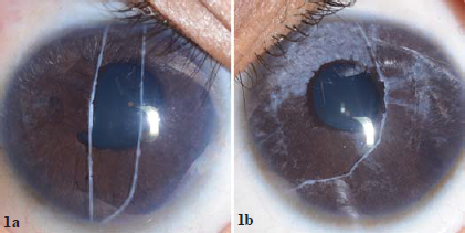
- Slit-lamp photograph with diffuse illumination showing megalocornea, a white, cord-like structure resting on the anterior surface of the iris representing detached Schwalbe line with attached iris strands, iris atrophy and ectropion uveae.
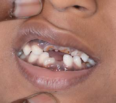
- Clinical photograph showing enamel hypoplasia.
An anteriorly displaced Schwalbe line or posterior embryotoxon with attached iris strands forms an essential component of the Axenfeld anomaly. Disordered neural crest cell migration and differentiation play an important role in the pathogenesis of Axenfeld–Rieger syndrome. The shared origin of Schwalbe line and iris stroma may explain the occurrence of detached Schwalbe line with attached iris strands. This distinctive finding has been reported sparsely in Axenfeld anomaly and Axenfeld–Rieger syndrome and represents a rare clinical sign of this disease spectrum.1,2
Conflicts of interest
None declared
References
- Detached Schwalbe's line in Axenfeld anomaly. Indian J Ophthalmol Case Rep. 2021;1:812-13.
- [CrossRef] [Google Scholar]
- Unusual presentation in Axenfeld-Rieger syndrome. Indian J Ophthalmol. 2011;59:312-14.
- [CrossRef] [Google Scholar]


