Generic selectors
Exact matches only
Search in title
Search in content
Post Type Selectors
Filter by Categories
Acknowledgements
Authors’ reply
Book Review
Book Reviews
Classics In Indian Medicine
Clinical Case Report
Clinical Case Reports
Clinical Research Methods
Clinico-pathological Conference
Clinicopathological Conference
Conferences
Correspondence
Corrigendum
Editorial
Eminent Indians in Medicine
Errata
Erratum
Everyday Practice
Film Review
History of Medicine
HOW TO DO IT
Images In Medicine
Indian Medical Institutions
Letter from Bristol
Letter from Chennai
Letter From Ganiyari
Letter from Glasgow
Letter from London
Letter from Mangalore
Letter From Mumbai
Letter From Nepal
Masala
Medical Education
Medical Ethics
Medicine and Society
News From Here And There
Notice of Retraction
Notices
Obituaries
Obituary
Original Article
Original Articles
Review Article
Selected Summaries
Selected Summary
Short Report
Short Reports
Speaking for Myself
Speaking for Ourselve
Speaking for Ourselves
Students@nmji
View/Download PDF

Translate this page into:

Images in Medicine
34 (
4
); 246-246
doi:
10.25259/NMJI_600_20

Double globes in single-orbit sign

Dr Rajendra Prasad Centre for Ophthalmic Sciences, All India Institute of Medical Sciences, New Delhi, India
Licence
This is an open-access article distributed under the terms of the Creative Commons Attribution-Non Commercial-Share Alike 4.0 License, which allows others to remix, tweak, and build upon the work non-commercially, as long as the author is credited and the new creations are licensed under the identical terms.

A 19-year-old man presented with complaints of outward displacement of the left eyeball for the past 2 years. On examination, the left eyeball had lateral dystopia of 6 mm. On T2-weighted MRI, a well-defined cystic lesion in the left orbit, resembling the other globe, was seen (Fig. 1). CT scan revealed a heterogeneous lesion in the left fronto-ethmoid sinus with extension into the orbit. A provisional diagnosis of left fronto-ethmoidal mucocoele was made. He underwent surgical removal of the mucocoele, and the affected ethmoid sinus was connected to the nasal cavity.
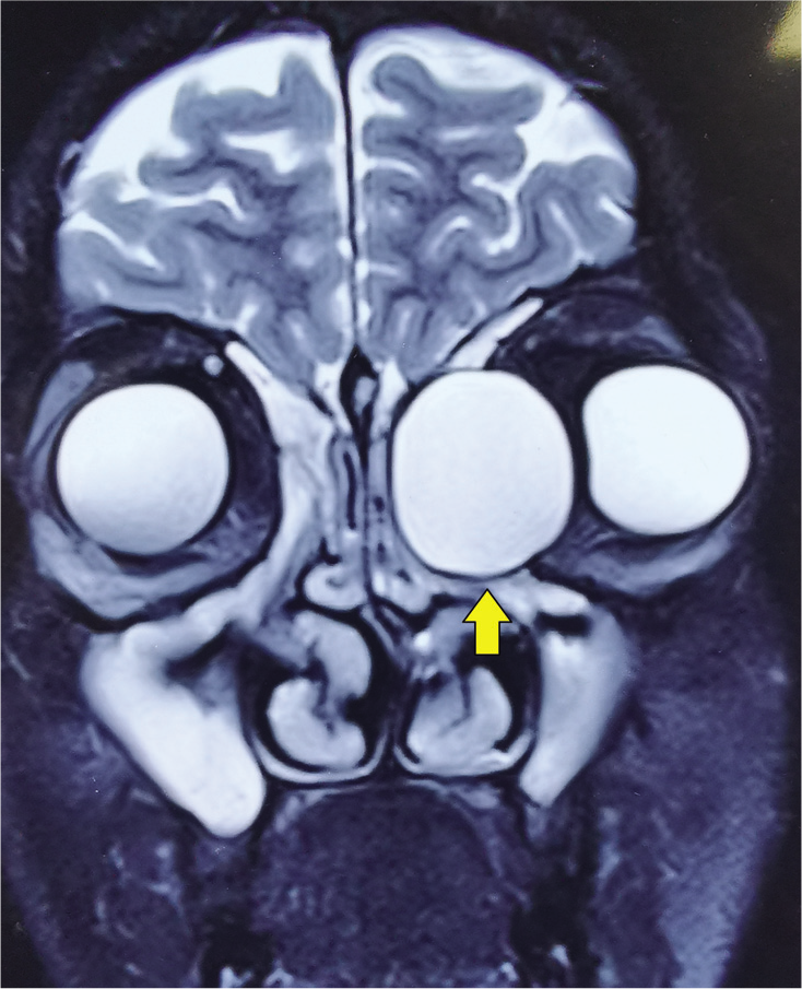
MRI showing a hyperintense cystic lesion on T2-weighted imaging at the left medial canthus, giving the appearance of ‘the double globe in single-orbit’ or ‘the third eyeball’ sign
FIG 1.
MRI showing a hyperintense cystic lesion on T2-weighted imaging at the left medial canthus, giving the appearance of ‘the double globe in single-orbit’ or ‘the third eyeball’ sign

Mucocoeles are epithelial-lined cystic lesions frequently located in the paranasal sinuses. Their pathogenesis remains a mystery and many believe the cause to be a sequelae of obstruction of the affected sinus due to chronic rhinosinusitis, trauma or surgery.1 The presenting symptoms usually include headache, recurrent episodes of nasal obstruction, diplopia, displacement of the ocular globe and in severe instances, meningitis.2 Mucocoeles in the fronto-ethmoid sinus displace the eyeball forwards, downwards and laterally. The investigation of choice is CT scan, as it provides details of the bony erosions and orbital and intracranial extensions. On T2-weighted MRI, these appear as well-defined cystic lesions and can mimic the globe, giving the appearance of double globes in a single orbit.3 Malignant lesions, dermoid cysts, fungal and tubercular granulomas and cholesterol granulomas form the list of differential diagnoses. The management of fronto-ethmoidal mucocoeles primarily involves surgical excision with marsupialization.4

Conflicts of interest

None declared

References

  1. . Image diagnosis: Frontoethmoidal mucocele. Perm J. 2019;23:18-288.
    [Google Scholar]
  2. , , . The natural history and clinical characteristics of paranasal sinus mucoceles: A clinical review. Int Forum Allergy Rhinol. 2013;3:712-17.
    [CrossRef] [PubMed] [Google Scholar]
  3. , , , , . Considerations in the management of giant frontal mucoceles with significant intracranial extension: A systematic review. Am J Rhinol Allergy. 2016;30:301-5.
    [CrossRef] [PubMed] [Google Scholar]
  4. , , . Contemporary management of frontal sinus mucoceles: A meta-analysis. Laryngoscope. 2014;124:378-86.
    [CrossRef] [PubMed] [Google Scholar]

Fulltext Views
1,655

PDF downloads
1,818
View/Download PDF
Download Citations
BibTeX
RIS
Show Sections