Translate this page into:
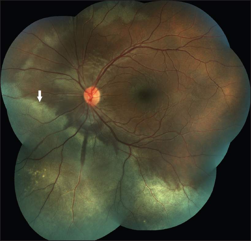
Extensive commotio retinae involving peripheral retina
Corresponding Author:
Rohan Chawla
Dr Rajendra Prasad Centre for Ophthalmic Sciences, All India Institute of Medical Sciences, New Delhi
India
dr.rohanrpc@gmail.com
| How to cite this article: Tripathy K, Chawla R. Extensive commotio retinae involving peripheral retina. Natl Med J India 2017;30:242 |
A 16-year-old boy was injured by a cricket ball a day before presenting to us. The left fundus showed large areas of whitish opacification of the peripheral retina ([Figure - 1], arrow) suggestive of commotio retinae. Visual acuity was 6/6 and the whiteness resolved at 1 week without any pigmentation.
 |
| Figure 1: Fundus photomontage of the left eye shows peripheral retinal whitening (commotio retinae, arrow) |
Following closed globe injury, commotio retinae occurs due to disruption of photoreceptor outer segments and is not true oedema. It may occur at the macula, manifesting as a cherry red spot (Berlin's oedema) and/or it can involve the peripheral retina. Commotio retinae commonly resolves spontaneously without sequelae. There is no known treatment.
Acknowledgement
We thank Trina Sengupta Tripathy for her support and technical assistance during preparation of the manuscript.
Fulltext Views
1,577
PDF downloads
714


