Translate this page into:
Gastric volvulus: Bird’s beak sign on computed tomography
[To cite: Kajihara Y. Gastric volvulus: Bird’s beak sign on computed tomography. Natl Med J India 2024;37:168. DOI: 10.25259/NMJI_330_2023]
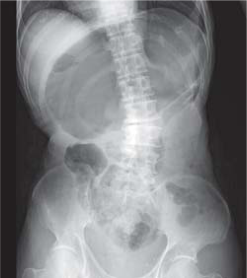
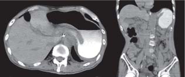
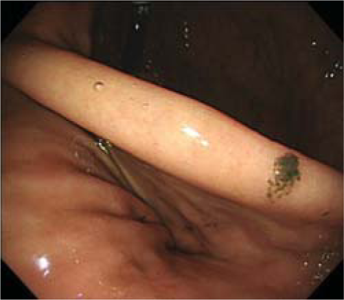
A 77-year-old man presented with acute onset of vomiting. His abdomen was distended, tender and tympanic. Abdominal X-ray showed massive gastric dilatation (Fig. 1). A nasogastric tube was inserted for decompression. In addition, computed tomography (CT) after administration of diatrizoate meglumine and diatrizoate sodium (Gastrografin®) showed displacement of the antrum above the gastro-oesophageal junction and progressive tapering of the stomach—the bird’s beak sign (Fig. 2). A diagnosis of gastric volvulus was made. In the present case gastric volvulus was considered to be primary since CT scan showed no splenic or diaphragmatic disorders. Gastroscopy confirmed twisting of the stomach without mucosal ischaemia (Fig. 3) and sequential endoscopic reduction was performed. He could take orally soon after successful reduction.

- Abdominal X-ray showing massive gastric dilatation

- Computed tomography after administration of Gastrografin® revealing displacement of the antrum above the gastro-oesophageal junction and progressive tapering of the stomach, known as the bird’s beak sign

- Gastroscopy confirming twisting of the stomach without mucosal ischaemia
The bird’s beak sign metaphorically describes the fluoroscopic appearance of sigmoid volvulus.1 However, this sign is also appreciated on CT images of gastric volvulus. Gastric volvulus can have a life-threatening clinical course due to ischaemia of the gastric mucosa.2 Therefore, prompt diagnosis and treatment are needed. Although it is difficult to determine the degree of mucosal ischaemia, CT is useful to demonstrate abnormal position and torsion of the stomach.3 For evaluating gastric mucosal ischaemia, early gastroscopy is recommended. Also sequential endoscopic reduction is a less-invasive and effective treatment.
Conflicts of Interest
None declared
References
- A review article on gastric volvulus: A challenge to diagnosis and management. Int J Surg. 2010;8:18-24.
- [CrossRef] [Google Scholar]
- The management of gastric volvulus in elderly patients. Int J Surg Case Rep. 2016;29:88-93.
- [CrossRef] [Google Scholar]


