
Translate this page into:
Idiopathic calcinosis cutis of scrotum
[To cite: Shaikh AS, Phatak SV, Dhok AP, Onkar PM, Dalvi VN. Idiopathic calcinosis cutis of scrotum. Natl Med J India 2024;37:169. DOI: 10.25259/NMJI_321_2023]
Idiopathic scrotal calcinosis is an uncommon, usually painless condition, characterised by one or more subcutaneous calcified nodules over the scrotum (Fig. 1). It is a benign condition but can cause problems in marital relationship.1 The nodules may involve any layer of the skin and progressively increase in size and number.2 The swellings consist of calcium and phosphorus deposits. Idiopathic scrotal calcinosis is predominantly seen in children but can also be seen in early adulthood. It occurs without any metabolic condition involving phosphorus and calcium metabolism. Calcinosis cutis is divided into four subtypes: Iatrogenic, dystrophic, metastatic and idiopathic.2 Idiopathic scrotal calcinosis has a scrotal skin with calcific deposits with surrounding granulomatous inflammation. Vulval calcinosis can occur in women.3 Histopathological examination is required because, clinically, it may be confused with onchocercoma, schwannoma, solitary neurofibroma, lipoma, steatoma and fibroma.4 Basophilic calcium deposits with surrounding inflammatory changes are seen on histopathological examination. Lesions which are small in size are treated with the pinch–punch excision whereas larger lesions need surgical excision.5
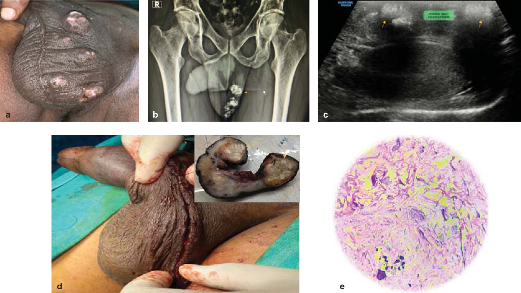
- Multiple whitish nodular swellings are seen over the scrotum on the left side (a). An X-ray of the pelvis and scrotum showed multiple calcific foci in the left hemi-scrotum (b). High frequency ultrasound of the scrotum showed multiple roughly oval hyperechoic lesions with posterior acoustic shadowing in the subcutaneous plane of the scrotum representing calcifications, both testes were normal (c). The postoperative photograph (d) after left hemiscrotectomy. Histopathological slide of the deeper dermis showed calcific foci separated by bands of collagenous tissue. The intervening fibrocollagenous connective tissue had lymphocytic inflammatory infiltrates (e)
Conflicts of interest
None declared
References
- Scrotal calcinosis effect––Case report and review of literature. Urol Case Rep. 2020;33:101347.
- [CrossRef] [Google Scholar]
- Idiopathic calcinosis cutis of the scrotum: A case report and review of the literature. J Med Case Rep. 2018;12:366.
- [CrossRef] [Google Scholar]
- Scrotal calcinosis: A case report and review of pathogenesis and surgical management. Case Rep Urol. 2012;2012:475246.
- [CrossRef] [Google Scholar]
- Calcinosis circumscripta of the scrotal wall: The etiological role of Onchocerca volvulus. Br J Dermatol. 1962;74:136-40.
- [CrossRef] [Google Scholar]
- Surgical pearl: Pinch–punch excisions for scrotal calcinosis. J Am Acad Dermatol. 2004;50:780-1.
- [CrossRef] [Google Scholar]


