Translate this page into:
Macrodystrophia lipomatosa: A rare form of localized gigantism
Corresponding Author:
Mohd Ilyas
Department of Radiodiagnosis, Sher-i-Kashmir Institute of Medical Sciences, Srinagar, Kashmir
India
ilyasmir40@gmail.com
| How to cite this article: Ilyas M, Ahmad Z, Choh N. Macrodystrophia lipomatosa: A rare form of localized gigantism. Natl Med J India 2020;33:250 |
An 11-year-old female was being evaluated for urinary obstruction by intravenous pyelography when the radiologist noticed her malformed right foot. On enquiry, the parents said it was since birth. They did not pay much attention to it. The patient had difficulty in walking for the past 2–3 years, but they had not consulted any doctor for the problem.
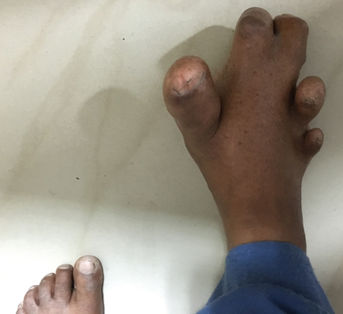
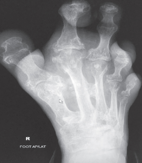
There was evidence of macrodactyly of the toes of the right foot with increased soft tissue compared to the normal left foot with abnormal position of the toes [Figure - 1]. A plain X-ray of the foot revealed marked hypertrophy of the first and second toes with soft tissue hypertrophy and splaying of the phalanges with fat translucencies in the soft tissue [Figure - 2]. A diagnosis of macrodystrophia lipomatosa was made and the patient counselled to visit an orthopaedic surgeon.
 |
| Figure 1: Photograph of the right foot showing gigantism involving the toes and soft tissues |
 |
| Figure 2: Anteroposterior plain X-ray of the right foot showing hypertrophied first and second toes with splaying of the phalanges and increased fat translucencies in the hypertrophied soft tissue suggesting macrodystrophia lipomatosa |
Macrodystrophia lipomatosa is a rare congenital localized gigantism of a limb or digit with progressive enlargement of the soft tissue components, especially the fibrofatty tissue.[1] The term was first coined by Feriz in 1925 for unilateral overgrowth of the lower limb.[2] It involves the foot more commonly than the hand or upper limb. It can be evaluated by ultrasonography, plain X-ray, CT scan or MRI. Microscopically, it is characterized by marked increase of all mesenchymal cells.[3] The mimickers include neurofibromatosis type 1, vascular malformations, neurofibrolipomatosis and Proteus syndrome. The mainstay of treatment remains surgery, which improves the cosmetic function with retained neurological function.[4]
Conflicts of interest. None declared
| 1. | Durairaj AR, Mahipathy SR. Macrodystrophia lipomatosa of the toe: A rare case report. J Clin Diagn Res 2016;10:PD27–8. [Google Scholar] |
| 2. | Feriz H. Makrodystrophia lipomatosa progressiva. Virchows Arch 1925;260:308–68. [Google Scholar] |
| 3. | Khan RA, Wahab S, Ahmad I, Chana RS. Macrodystrophia lipomatosa: Four case reports. Ital J Pediatr 2010;36:69. [Google Scholar] |
| 4. | Brodwater BK, Major NM, Goldner RD, Layfield LJ. Macrodystrophia lipomatosa with associated fibrolipomatous hamartoma of the median nerve. Pediatr Surg Int 2000;16:216–18. [Google Scholar] |
Fulltext Views
2,108
PDF downloads
2,065


