Generic selectors
Exact matches only
Search in title
Search in content
Post Type Selectors
Filter by Categories
Acknowledgements
Authors’ reply
Book Review
Book Reviews
Classics In Indian Medicine
Clinical Case Report
Clinical Case Reports
Clinical Research Methods
Clinico-pathological Conference
Clinicopathological Conference
Conferences
Correspondence
Corrigendum
Editorial
Eminent Indians in Medicine
Errata
Erratum
Everyday Practice
Film Review
History of Medicine
HOW TO DO IT
Images In Medicine
Indian Medical Institutions
Letter from Bristol
Letter from Chennai
Letter From Ganiyari
Letter from Glasgow
Letter from London
Letter from Mangalore
Letter From Mumbai
Letter From Nepal
Masala
Medical Education
Medical Ethics
Medicine and Society
News From Here And There
Notice of Retraction
Notices
Obituaries
Obituary
Original Article
Original Articles
Review Article
Selected Summaries
Selected Summary
Short Report
Short Reports
Speaking for Myself
Speaking for Ourselve
Speaking for Ourselves
Students@nmji
View/Download PDF

Translate this page into:

Images in Medicine
36 (
1
); 55-55
doi:
10.25259/NMJI_99_22

Metastatic calcification: An uncommon issue in peritoneal dialysis

Department of Paediatrics, All India Institute of Medical Sciences, Bilaspur, India
VPO Bela Teh Nadaun, Hamirpur, India
Department of Nephrology, Indira Gandhi Medical College and Hospital, Shimla Himachal Pradesh, India
Licence
This is an open-access article distributed under the terms of the Creative Commons Attribution-Non Commercial-Share Alike 4.0 License, which allows others to remix, tweak, and build upon the work non-commercially, as long as the author is credited and the new creations are licensed under the identical terms.

[To cite: Gupta S, Sadan P, Singh R, Kumar A. Metastatic calcification: An uncommon issue in peritoneal dialysis. Natl Med J India 2023;36:55. DOI: 10.25259/NMJI_99_22]

A 64-year-old woman, diagnosed with chronic kidney disease, stage 5 and on peritoneal dialysis (CKD 5 PD) for the past 3 years, presented with pain in the right shoulder. Her PD schedule comprised of three exchanges of 1.5% and one of 7.5% dialysate (Baxter, India) with 3.5 mEq/L calcium. She was on intermittent calcium carbonate and phosphate binder, and calcitriol. The patient was anuric with ultrafiltration of 1200 ml/day. She did not have any history of skin rash, oral ulcer and colaour changes in her extremities or trauma. On examination, tbhe patient had swelling and a limited range of motion at the right shoulder joint. Investigations revealed serum calcium 13.3 mg/dl, serum phosphorus 4.3 mg/dl, 25-hydroxyvitamin D 34.30 ng/ml and intact parathyroid hormone level 19.2 pg/ml. X-ray of the joint showed well-circumscribed, multilobular, calcified periarticular radio-opacity without involvement of bones, suggestive of tumoural calcinosis (Fig. 1a). Following this, calcium supplements and calcitriol were stopped and cholecalciferol was added. The patient was continued on PD with the same composition because of non-availability of low calcium dialysate. After 6 months of treatment, there was a marked improvement in symptoms, serum calcium (8.9 mg/dl) and X-ray findings (Fig. 1b).
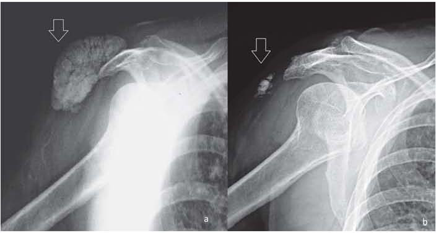
X-ray of the right shoulder joint: (a) white arrow showing massive calcium deposition, which is well-circumscribed, multilobular, periarticular without involvement of the bones; (b) residual calcium deposits after treatment
FIG 1.
X-ray of the right shoulder joint: (a) white arrow showing massive calcium deposition, which is well-circumscribed, multilobular, periarticular without involvement of the bones; (b) residual calcium deposits after treatment

Metastatic calcification predominately affects the kidneys, lungs, stomach and the media of arteries, but cutaneous and subcutaneous tissues may be involved.1 Predisposing factors include an increase in calcium–phosphorus in the serum, the degree of secondary hyperparathyroidism, the level of blood magnesium, the degree of alkalosis, and the presence of local tissue injury.2

Conflicts of interest

None declared

References

  1. . Metastatic calcification of the hand in a patient undergoing hemodialysis. Am J Med. 2004;116:572-3.
    [CrossRef] [PubMed] [Google Scholar]
  2. , , , . Benign nodular calcification and calciphylaxis in a haemodialysed patient. J Eur Acad Dermatol Veneraeol. 1998;11:69-71.
    [CrossRef] [Google Scholar]

Fulltext Views
1,005

PDF downloads
876
View/Download PDF
Download Citations
BibTeX
RIS
Show Sections