Generic selectors
Exact matches only
Search in title
Search in content
Post Type Selectors
Filter by Categories
Acknowledgements
Authors’ reply
Book Review
Book Reviews
Classics In Indian Medicine
Clinical Case Report
Clinical Case Reports
Clinical Research Methods
Clinico-pathological Conference
Clinicopathological Conference
Conferences
Correspondence
Corrigendum
Editorial
Eminent Indians in Medicine
Errata
Erratum
Everyday Practice
Film Review
History of Medicine
HOW TO DO IT
Images In Medicine
Indian Medical Institutions
Letter from Bristol
Letter from Chennai
Letter From Ganiyari
Letter from Glasgow
Letter from London
Letter from Mangalore
Letter From Mumbai
Letter From Nepal
Masala
Medical Education
Medical Ethics
Medicine and Society
News From Here And There
Notice of Retraction
Notices
Obituaries
Obituary
Original Article
Original Articles
Review Article
Selected Summaries
Selected Summary
Short Report
Short Reports
Speaking for Myself
Speaking for Ourselve
Speaking for Ourselves
Students@nmji
View/Download PDF

Translate this page into:

Images in Medicine
36 (
2
); 133-133
doi:
10.25259/NMJI_276_22

Tracheal tumorous tuberculosis

Department of Pulmonary Medicine, All India Institute of Medical Sciences, Ansari Nagar, New Delhi, India
Licence
This is an open-access article distributed under the terms of the Creative Commons Attribution-Non Commercial-Share Alike 4.0 License, which allows others to remix, tweak, and build upon the work non-commercially, as long as the author is credited and the new creations are licensed under the identical terms.

[To cite: Mittal S, Mohan A, Madan K, Hadda V, Guleria R. Tracheal tumorous tuberculosis. Natl Med J India 2023;36:133. DOI: 10.25259/NMJI_276_22]

A 74-year-old, non-smoker man presented with shortness of breath and cough for 2 months. There was no history of fever, haemoptysis or chest pain. He had no peripheral lymphadenopathy and his vital signs were within normal limits. His liver and renal function tests, urinalysis, electrocardiogram and a posteroanterior chest X-ray were unremarkable. CT scan of the thorax showed a tumorous lesion in the mid-trachea with clear lung fields (Fig. 1a). On bronchoscopic examination, a broad-based lesion was seen arising from the posterior wall of the trachea causing 30%–40% luminal narrowing (Fig. 1b). The rest of the bronchial tree was normal. An electrosurgical snaring of the mass was done to avoid airway bleeding during the biopsy and to achieve airway recanalization (Fig. 1c). The histopathological examination revealed epithelioid cell granulomas with focal necrosis along with few acid-fast bacilli suggesting a diagnosis of tumorous endobronchial tuberculosis and the MTBRif assay of the tissue detected Mycobacterium tuberculosis (Fig. 2). Antitubercular therapy was initiated, and follow-up bronchoscopy after 6 months of therapy showed a scar-like area in the posterior wall of the trachea.

FIG 1.
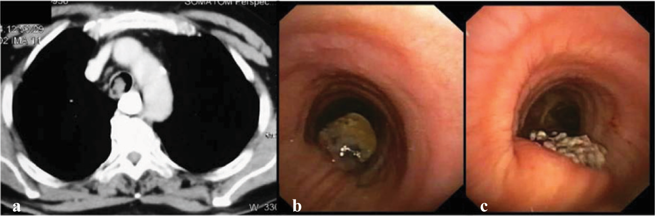
(a) CT thorax showing an endotracheal mass; (b) bronchoscopic appearance of the mass in the posterior wall of the trachea; (c) post-snaring view of the posterior wall of the trachea
FIG 2.
Microphotograph of the tracheal mass: (a) surface squamous metaplasia (red arrow) and the underlying submucosa showing many well-formed epithelial cell granulomas with Langhans type of giant cell (yellow arrow); (b) acid-fast stain shows few dispersed viable acid-fast bacilli morphology consistent with Mycobacterium tuberculosis

Endobronchial involvement is uncommon in tuberculosis. Endobronchial tuberculosis is of various types including caseating, oedematous–hyperaemic, fibro-stenotic, tumorous, granular, ulcerative and non-specific bronchitis.1 The tumorous type is extremely rare and is often initially misdiagnosed as carcinoma.2

Conflicts of interest

None declared

References

  1. , , , , , . Bilateral multiple tumor-like endobronchial tuberculosis, diagnosed with bronchoscopic examination. Tuberk Toraks. 2011;59:266-70.
    [CrossRef] [Google Scholar]
  2. , , . Endobronchial tuberculosis simulating lung cancer. Indian J Chest Dis Allied Sci. 1997;39:251-4.
    [Google Scholar]

Fulltext Views
783

PDF downloads
6,351
View/Download PDF
Download Citations
BibTeX
RIS
Show Sections