
Translate this page into:
Ultra-wide-field fundus autofluorescence and asteroid hyalosis
riksag@gmail.com
How to cite this article: MISHRA C, SEN S, KANNAN NB, RAMASAMY K. Ultra-wide-field fundus autofluorescence and asteroid hyalosis. Natl Med J India 34:2021;186.
Asteroid hyalosis (AH) may lead to a media haze, which persists on fundus photography. Ultra-wide-field fundus autofluorescence (FAF) helps in viewing the posterior pole in eyes with dense AH and enables the surgeon to assess the adequacy of laser photocoagulation.
AH is a degenerative condition of the vitreous due to collection of calcium globules, associated with the presence of systemic diseases such as diabetes mellitus, hypertension and hyperlipidaemia.1 Often, AH can cause considerable media haze and prevent ophthalmologists from viewing the posterior pole of the eye to assess underlying retinal diseases. Bilateral AH may be seen commonly in the population with diabetes.2 Previous reports have estimated that 8.5% of ungradable images can be associated with AH.3 We present two such patients of proliferative diabetic retinopathy (PDR) with extensive AH (Figs 1 and 2). In patient 1 (Fig. 1), the left eye had media haze due to AH, and on FAF, we could determine the adequacy of panretinal photocoagulation (PRP), following which the patient was advised observation. In patient 2 (Fig. 2), both eyes showed media haze, which cleared on FAF, and additional photocoagulation was advised for both eyes. We evaluated the patients using FAF on Optos Daytona Plus (Optos PLC, Dunfermline, UK), which projects green light of 532 nm wavelength and detects emitted signals in the range of 570–580 nm. Conventional slit-lamp biomicroscopy uses pan-fundoscopic lenses or colour. Since the entirety of emitted light originates from the retina and is detected by a pre-calibrated detector in the range of 570–580 nm, a ‘single pass’ of light occurs. The light reflected from the asteroid bodies scatters back towards the retina and does not reduce the captured FAF image quality.4 Thus, FAF is a valuable non-invasive tool for the treatment of PDR and also as a teaching tool.

- Optos ultra-wide-field pseudocolour images of the right and left eyes of patient 1 with proliferative diabetic retinopathy with asteroid hyalosis. On fundus autofluorescence, the posterior pole of the left eye is visible
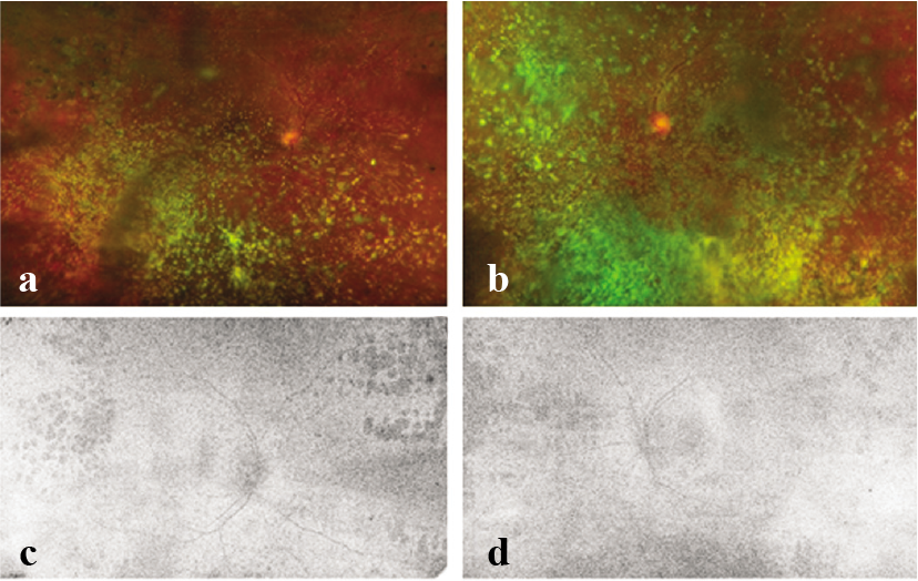
- Optos ultra-wide-field pseudocolour images of the right and left eyes of patient 2 with proliferative diabetic retinopathy with asteroid hyalosis. On fundus autofluorescence, the posterior poles of both eyes are visible. Patient 2 was advised additional laser photocoagulation in both eyes
Conflicts of interest
None declared
References
- Asteroid hyalosis: Pathogenesis and prospects for prevention. Eye (Lond). 2008;22:1278-85.
- [CrossRef] [PubMed] [Google Scholar]
- Frequency of diabetics in asteroid hyalosis patients. J Ayub Med Coll Abbottabad. 2003;15:10-11.
- [Google Scholar]
- Asteroid hyalosis and photographic diabetic retinopathy screening. Diabet Med. 2010;27:1093.
- [CrossRef] [PubMed] [Google Scholar]
- Photocoagulation guided by wide-field fundus autofluorescence in eyes with asteroid hyalosis. Eye (Lond). 2014;28:634-5.
- [CrossRef] [PubMed] [Google Scholar]


