Generic selectors
Exact matches only
Search in title
Search in content
Post Type Selectors
Filter by Categories
Acknowledgements
Authors’ reply
Book Review
Book Reviews
Classics In Indian Medicine
Clinical Case Report
Clinical Case Reports
Clinical Research Methods
Clinico-pathological Conference
Clinicopathological Conference
Conferences
Correspondence
Corrigendum
Editorial
Eminent Indians in Medicine
Errata
Erratum
Everyday Practice
Film Review
History of Medicine
HOW TO DO IT
Images In Medicine
Indian Medical Institutions
Letter from Bristol
Letter from Chennai
Letter From Ganiyari
Letter from Glasgow
Letter from London
Letter from Mangalore
Letter From Mumbai
Letter From Nepal
Masala
Medical Education
Medical Ethics
Medicine and Society
News From Here And There
Notice of Retraction
Notices
Obituaries
Obituary
Original Article
Original Articles
Review Article
Selected Summaries
Selected Summary
Short Report
Short Reports
Speaking for Myself
Speaking for Ourselve
Speaking for Ourselves
Students@nmji
View/Download PDF

Translate this page into:

Images in Medicine
34 (
6
); 373-373
doi:
10.25259/NMJI_57_21

Vortex vein ampullae seen in oculocutaneous albinism on ultrawide field imaging without angiography

Department of Vitreo-Retinal Services Aravind Eye Hospital and Post Graduate Institute of Ophthalmology Madurai, Tamil Nadu, India
Licence
This is an open-access article distributed under the terms of the Creative Commons Attribution-Non Commercial-Share Alike 4.0 License, which allows others to remix, tweak, and build upon the work non-commercially, as long as the author is credited and the new creations are licensed under the identical terms.

To cite: Mishra C, Kohli P, Babu N. Vortex vein ampullae seen in oculocutaneous albinism on ultrawide field imaging without angiography. Natl Med J India 2021;34:373.

A 27-year-old man presented with defective vision and nystagmus since childhood in both eyes. His best-corrected vision was 20/80 in both eyes, with a refractive error of –2DS. Anterior segment examination showed a transillumination iris defect. Posterior segment examination showed hypopigmented albinotic fundus with prominent background choroidal vasculature, multiple vortex vein ampullae near the equator and dilated posterior vortex vein forming ampulla in the peripapillary region (Fig. 1). He was diagnosed as oculocutaneous albinism with moderate myopia. He was advised spectacles and counselled about low-vision aids.
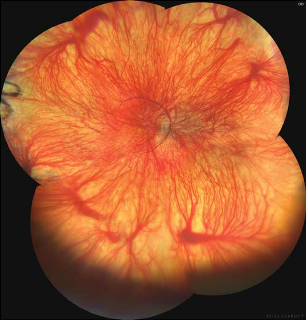
Ultrawide field image (Clarus 500, Carl Zeiss Meditech, Dublin, USA) showing hypopigmented albinotic fundus with prominent background choroidal vasculature, multiple vortex vein ampullae near the equator and dilated posterior vortex vein forming ampulla in the peripapillary region
FIG 1.
Ultrawide field image (Clarus 500, Carl Zeiss Meditech, Dublin, USA) showing hypopigmented albinotic fundus with prominent background choroidal vasculature, multiple vortex vein ampullae near the equator and dilated posterior vortex vein forming ampulla in the peripapillary region

Although indocyanine green angiography is usually required to visualize the vortex vein ampullae, they can sometimes be visualized on fundoscopy in an albinotic fundus.1,2 Wide-angle imaging systems can be used to document these ampullae.2

Conflicts of interest

None declared.

References

  1. , , , . Detection of posterior vortex veins in eyes with pathologic myopia by ultra-widefield indocyanine green angiography. Br J Ophthalmol. 2017;101:1179-84.
    [CrossRef] [PubMed] [Google Scholar]
  2. , . Choroidal vasculature without angiography. Indian J Ophthalmol. 2019;67:141.
    [CrossRef] [PubMed] [Google Scholar]

Fulltext Views
1,947

PDF downloads
1,170
View/Download PDF
Download Citations
BibTeX
RIS
Show Sections