
Translate this page into:
Bilateral orbital cellulitis in an infant: An unusual case
Correspondence to SIDDHARTH MADAN; drsiddharthmadan@gmail.com
To cite: Madan S, Beri S, Virmani P, Khan S, Chakravarti A. Bilateral orbital cellulitis in an infant: An unusual case. Natl Med J India 2021;34:213–15.
Abstract
Bilateral orbital cellulitis is an uncommon presentation in infants, and its association with rhinosinusitis has been scantily reported in the literature. An infant underwent bilateral uncinectomy and right ethmoidal decompression for bilateral orbital cellulitis with right ethmoidal sinusitis, after a period of non-recovery with conservative treatment. Mixed infection with Escherichia coli and methicillin-sensitive Staphylococcus aureus was observed. The right eyelid swelling began to resolve; however, the left lower eyelid showed an increase in the fluctuant swelling. Transconjunctival incision and drainage of pus was done in the left eye subsequently. The patient showed marked clinical recovery and is doing well. A multidisciplinary management approach can avert potentially life-threatening sequelae of this condition.
INTRODUCTION
Orbital cellulitis is a relatively uncommon inflammatory process involving structures anterior to the orbital septum, and is more common in children. The most common manifestations are erythema and oedema of eyelids, external ophthalmoplegia, proptosis, conjunctival chemosis and reduced visual acuity. The common predisposing factors are sinus infection, eyelid infection, dental abscess or haematogenous spread. A history of upper respiratory tract infection is important in children. Early diagnosis and management is necessary to avoid serious complications such as intracranial abscess, meningitis, cavernous sinus thrombosis, vision loss, sepsis and even death. We report an infant in whom medical and surgical interventions led to complete resolution.
THE CASE
A 2.5-month-old child presented to the ophthalmology outpatient department (OPD) with complaints of swelling, redness and purulent discharge in her right eye for 6 days and similar complaints in her left eye for 2 days (Fig. 1a–c). The swelling was associated with fever and nasal discharge. There was neither any history of upper respiratory tract infection before the commencement of swelling in the eyes nor any history of protrusion of either eye, trauma, ear discharge, cough, anorexia, weight loss, vesicular lesions, vomiting or seizures. Two days following the onset of complaints, the parents consulted a general physician who advised instillation of topical eye drops. Apparently, there was no relief. The child was seen by another paediatrician 2 days later who started oral amoxicillin and clavulanic acid and advised magnetic resonance imaging (MRI) of the orbits. The child was referred to a higher centre for opinion. Two days later, the child presented to the ophthalmology OPD. Examination revealed diffuse erythematous swelling involving both upper and lower eyelids in the right eye and only the lower eyelid in the left eye. She was unable to open her right eye. There was grade 2 (marked) limitation of ocular movements. Conjunctival chemosis and congestion along with purulent discharge was present in both the eyes. Although the right eye seemed to have mild proptosis, comprehensive clinical examination of proptosis was limited by the presence of extensive eyelid swelling. Fundus and pupil reactions were normal bilaterally. Considering a working diagnosis of orbital cellulitis, the child was admitted. The possibility of cavernous sinus thrombosis is likely in such presentations; however, demonstration of normal pupil reactions and preserved abduction in the left eye suggested its absence.
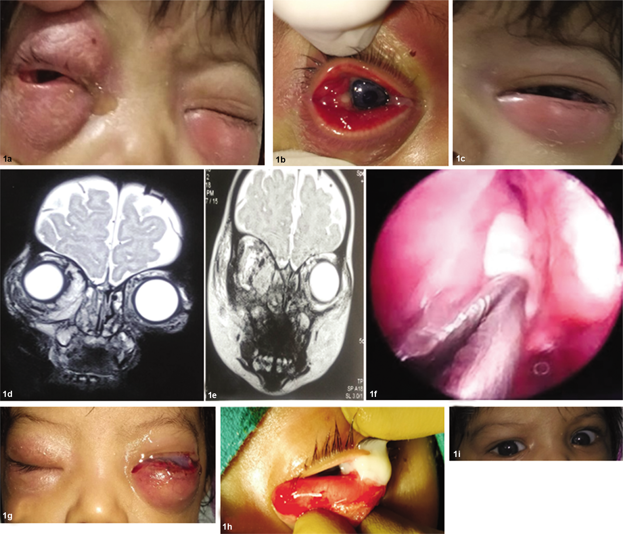
- Clinical presentation of the patient with radiological imaging and intraoperative photographs. Presentation with swelling, redness and purulent discharge in the right eye (a and b) and similar complaints in the left eye (c). Magnetic resonance imaging showed both eye orbital cellulitis and right side ethmoidal sinusitis (d and e). Bilateral uncinectomy and right ethmoidal decompression performed under general anaesthesia (f). Right eyelid oedema decreased considerably, left eyelid oedema increased considerably (g). Left eye transconjunctival incision and drainage performed under general anaesthesia (h). One month following complete recovery (i)
She was started on intravenous antibiotics (amoxicillin with clavulanic acid and amikacin) with fortified tobramycin (1.4%) eye drops hourly. Laboratory investigations showed a raised C-reactive protein and total leucocyte count. The MRI showed features of orbital cellulitis in both eyes and ethmoidal sinusitis of the right side (Fig. 1d and e). Microbiological analysis of the conjunctival swab sent for culture studies was not available at this time. Based on an otorhinolaryngology opinion, the patient had bilateral uncinectomy and right ethmoidal decompression under general anaesthesia (Fig. 1f). Four millilitres of pus was drained and sent for culture. Methicillin-sensitive Staphylococcus aureus and Escherichia coli were grown from this pus. E. coli was sensitive to ceftazidime and fortified ceftazidime eye drops were also given on an hourly basis. In the next 2 days, her right eyelid oedema decreased substantially. However, the left eyelid oedema increased (Fig. 1g). Hence, transconjunctival incision and drainage of the pus from the left eye was done under general anaesthesia (Fig. 1h). Based on the culture and antibiotic sensitivity report, the patient was started on intravenous ceftriaxone 210 mg b.d. for 8 days and vancomycin 6 hourly for 12 days along with the fortified eye drops (ceftazidime and tobramycin). The eyelid oedema gradually decreased and resolved completely by day 18 of admission. Refraction was done 1 month after recovery (Fig. 1i) and revealed +2.00 dioptre sphere of hypermetropia. She was fully immunized for age as per the National Immunisation Schedule for India and had received her first immunization dose scheduled at 6 weeks after birth for Haemophilus influenzae as a part of a pentavalent vaccine and also for Streptococcus pneumoniae. The child is on follow-up and has received her 10th- and 14th-week dosage of H. influenzae and S. pneumoniae vaccine, respectively. She is doing well.
DISCUSSION
Orbital cellulitis is most commonly associated with sinusitis. Bacterial orbital cellulitis attributable to sinusitis is primarily a disease of children although it can affect any age group.1 Acute bacterial sinusitis is present in 60%–80% of cases.2,3 The spread of infection from the sinuses to the orbit by direct invasion or haematogenous spread is facilitated by the relatively thin orbital bones and valveless venous connection between the orbit and the ethmoidal sinus.4 Ethmoidal sinus is the most common site to be involved as in our case, followed by the maxillary sinus. Bacteria usually associated with orbital cellulitis include S. aureus, Staphylococcus epidermidis, streptococci, especially S. pneumoniae, diphtheroids, H. influenzae, E. coli and also multiple species comprising both aerobes and anaerobes.5 Extraintestinal pathogenic E. coli (ExPEC) have considerable diversity in their genome and have a wide range of virulence-associated factors including toxins, adhesions, lipopolysaccharides, polysaccharide capsules, proteases and invasins, all of which are encoded by genes.6 These factors contribute to fitness of ExPEC and increase the adaptability and their ability to infect and proliferate inside the human body.6
Antimicrobial resistance to multiple drugs in E. coli has increased worldwide with high levels of resistance to ampicillin, amoxicillin–clavulanic acid (74.4%), norfloxacin, cefuroxime and ceftriaxone (71.4%).7 The pattern of susceptibility of E. coli to antibiotics shows a major geographical variation.8 Cephalosporin may be useful in the treatment of infections due to E. coli as it worked well in our patient.9 Blood culture may not be helpful in the diagnosis of orbital cellulitis as the pathology is often localized. Radiological investigations help in asssessing the extent of orbital cellulitis. Sonography offers the advantage that it is portable, provides a quick assessment and does not need anaesthesia in children.10 It can also be repeated as and when required. As this child presented to us with a MRI, we did not do an orbital ultrasound. Her follow-up examination with orbital sonography showed clinical recovery. MRI provides a dependable differentiation between various appearances of orbital inflammation.11 Medical therapy with broad spectrum i.v. antibiotics should be initiated as soon as the diagnosis of orbital cellulitis is suspected.12 Surgical treatment is required if there is underlying sinus disease, orbital or subperiosteal abscess or both especially in children, if there is no response to the antibiotics, there is progressive decrease in vision, or there is an orbital foreign body. Drainage of both the sinuses and the orbit should preferably be facilitated by both ophthalmological and otorhinolaryngological approaches. Functional endoscopic sinus surgery is performed for the drainage of sinus, and an 18G cannula allows easy drainage of the orbit.
Our patient, a 2.5-month-old infant, was younger compared with the study by Singh et al.13 where the average age of presentation was 5 years. Skedros et al. reviewed 30 children with subperiosteal orbital abscess who were 2 to 14 years of age.14 Caversaccio et al. in 2005 reported the earliest age of presentation at 5 weeks after birth. The hospital stay was 18 days in our patient and was consistent with the literature.13,15 One eye was involved more than the other, as has been reported in the literature.13 Bilateral orbital cellulitis after rhinosinusitis is a rare manifestation. Mitchell et al. in 2002 reported three children with bilateral orbital cellulitis as a complication of rhinosinusitis. It explains direct spread of the disease from the ethmoid sinuses to both orbits, without intracranial spread of infection.16 Thus, bilateral orbital cellulitis need not be a sequelae of cavernous sinus thrombosis as it can occur after ethmoidal sinusitis.
Conflicts of interest
None declared
References
- The acute orbit, Preseptal (periorbital) cellulitis, subperiosteal abscess and orbital cellulitis due to sinusitis. J Laryngol Otol. 1987;12(Suppl):1-8.
- [Google Scholar]
- Orbital cellulitis demands early recognition, urgent admission and aggressive management. J Accid Emerg Med. 1995;12:151-3.
- [CrossRef] [PubMed] [Google Scholar]
- 'The pathogenesis of orbital complications in acute sinusitis' (Laryngoscope 1970.801414-28) Laryngoscope. 1997;107:441-6.
- [CrossRef] [PubMed] [Google Scholar]
- The hot orbit: Orbital cellulitis. Middle East Afr J Ophthalmol. 2012;19:34-42.
- [CrossRef] [PubMed] [Google Scholar]
- Extraintestinal pathogenic Escherichia coli: An update on antimicrobial resistance, laboratory diagnosis and treatment. Expert Rev Anti Infect Ther. 2012;10:1165-76.
- [CrossRef] [PubMed] [Google Scholar]
- Antimicrobial resistance pattern in Escherichia coli causing urinary tract infection among inpatients. Indian J Med Res. 2014;139:945-8.
- [Google Scholar]
- Antimicrobial resistance of Escherichia coli and therapeutic implications. Int J Med Microbiol. 2005;295:503-11.
- [CrossRef] [PubMed] [Google Scholar]
- Reduced rates of antimicrobial resistance in Staphylococcus intermedius group and Escherichia coli isolated from diseased companion animals in an animal hospital after restriction of antimicrobial use. J Infect Chemother. 2019;25:531-6.
- [CrossRef] [PubMed] [Google Scholar]
- Using orbital sonography to diagnose and monitor treatment of acute swelling of the eyelids in pediatric patients. AJR Am J Roentgenol. 2002;179:1529-34.
- [CrossRef] [PubMed] [Google Scholar]
- MR imaging and CT of orbital infections and complications in acute rhinosinusitis. Radiol Clin North Am. 1998;36:1165-83, xi
- [CrossRef] [Google Scholar]
- Periorbital and orbital cellulitis in the Haemophilus influenzae vaccine era. J Pediatr Ophthalmol Strabismus. 1997;34:293-6.
- [CrossRef] [Google Scholar]
- Bilateral orbital complications of paediatric rhinosinusitis. Med J Armed Forces India. 2014;70:68-72.
- [CrossRef] [PubMed] [Google Scholar]
- Subperiosteal orbital abscess in children: Diagnosis, microbiology, and management. Laryngoscope. 1993;103:28-32.
- [CrossRef] [PubMed] [Google Scholar]
- Orbital complications of acute pediatric rhinosinusitis: Medical treatment versus surgery and analysis of the computer tomogram. Laryngorhinootologie. 2005;84:817-21.
- [CrossRef] [PubMed] [Google Scholar]
- Bilateral orbital complications of pediatric rhinosinusitis. Arch Otolaryngol Head Neck Surg. 2002;128:971-4.
- [CrossRef] [PubMed] [Google Scholar]


