
Translate this page into:
Choroidal tubercles
[To cite: Banerjee M, Kaur I, Venkatesh P. Choroidal tubercles. Natl Med J India 2023;36:338. DOI: 10.25259/NMJI_763_2022]
A 33-year-old woman with diagnosed pulmonary tuberculosis on antitubercular treatment presented with diminution of vision in the left eye. A subretinal healed tubercle inferotemporal to the disc was seen OD with outer retinal defect, intact retinal pigment epithelium (RPE), and enhanced transmission defect at the region of the tubercle (Figs 1a, c). Multiple healed choroidal tubercles were seen OS (Fig. 1b). The tubercle along inferotemporal arcade is surrounded by subretinal fluid involving the macula. A neurosensory detachment at the fovea along with intraretinal cystic spaces, subretinal fluid, hyperreflective inflammatory infiltrate above the RPE with loss of outer retinal layer details, intact RPE, and increased transmission signal can be seen at the area of the inferotemporal healed tubercle (Fig. 1d). Swept-source optical coherence tomography (OCT) may serve as an important adjunct in the follow up to monitor response to treatment and further management of complications such as choroidal neovascular membrane formation.
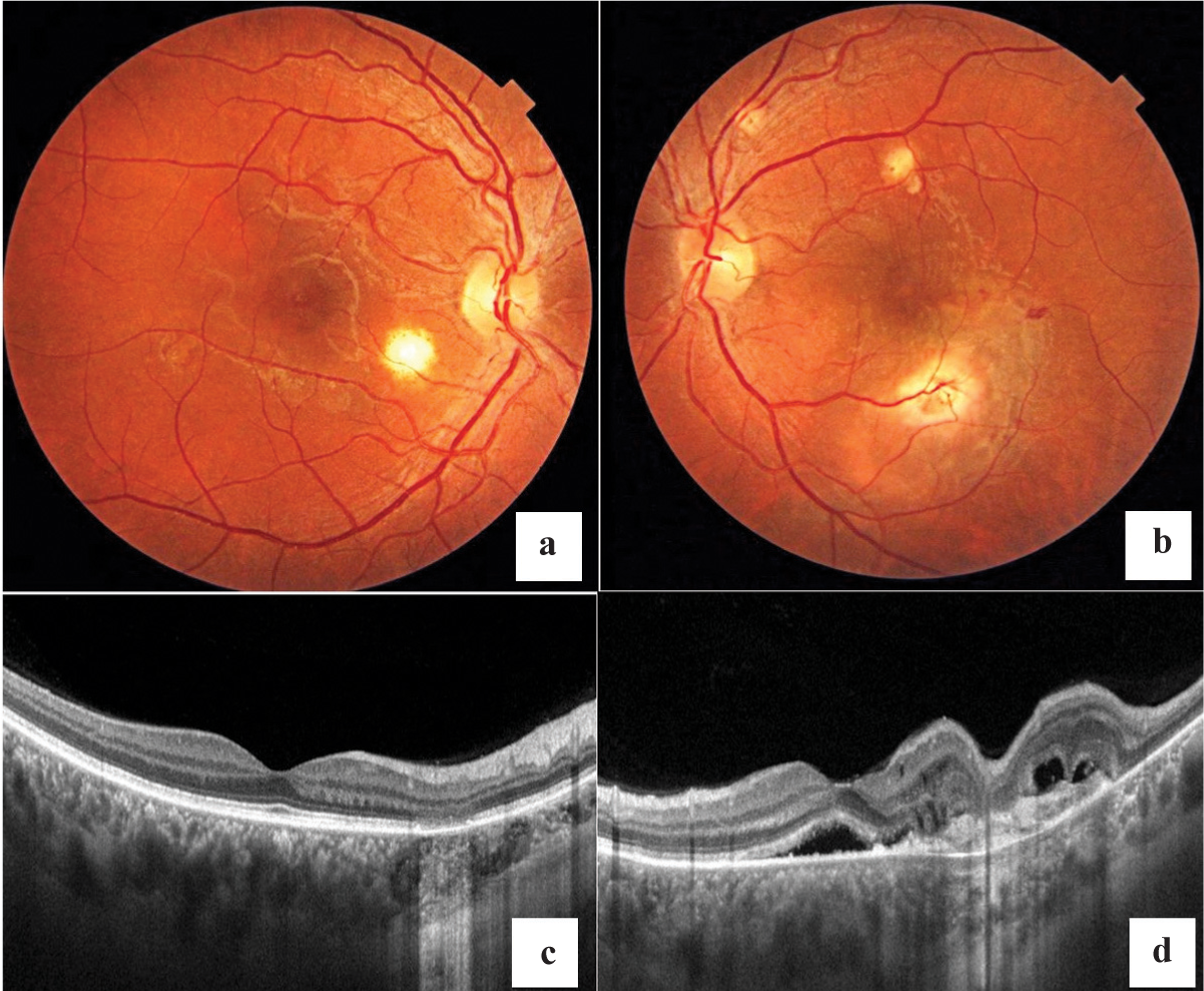
- Clinical photograph of the right eye reveals a subretinal healed tubercle inferotemporal to the disc (a) and multiple healed choroidal tubercles in the left eye (b). Swept-source optical coherence tomography (OCT) through the tubercle reveals outer retinal defect, intact retinal pigment epithelium (RPE), and enhanced transmission defect in the right eye (c) and a neuro-sensory detachment at the fovea along with intraretinal cystic spaces, subretinal fluid, hyperreflective inflammatory infiltrate above the RPE with loss of outer retinal layer details, intact RPE, and increased transmission signal at the area of the inferotemporal healed tubercle in the left eye (d)
Conflicts of interest
None declared


