Translate this page into:
Ectopia lentis and blue sclera in hyperhomocysteinaemia
Corresponding Author:
Brijesh Takkar
Dr Rajendra Prasad Centre for Ophthalmic Sciences, All India Institute of Medical Sciences, New Delhi
India
britak.aiims@gmail.com
| How to cite this article: Rathi A, Takkar B, Azad S. Ectopia lentis and blue sclera in hyperhomocysteinaemia. Natl Med J India 2017;30:176 |
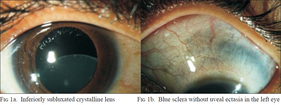
An 8-year-old girl was detected to have corrected visual acuity of 6/18 in both eyes during a school screening programme. Both her eyes had inferiorly subluxated lenses, the superior border of the lenses lying centrally within the visual axis [Figure - 1]a. There was an additional patch of blue discoloration of the sclera without uveal ectasia in the left eye [Figure - 1]b. A diagnostic work-up for bilateral ectopia lentis revealed high serum homocysteine levels. Her parents were counselled for lensectomy on follow-up, advised a consultation with a paediatrician and initiation of pyridoxine therapy.
 |
| Figure 1: |
The ligaments responsible for keeping the lens in position and shape are abundant in cysteine, and deficiency of the same due to homocystinuria results in ectopia lentis. Blue sclera is typically associated with other causes of ectopia lentis affecting the collagen structure and is uncommon in homocystinuria. A simple torchlight examination of the eyes can therefore provide valuable clues towards a serious underlying metabolic disorder.
Fulltext Views
1,734
PDF downloads
2,062


