Translate this page into:
Hemispheric cerebral oedema due to intracranial dural arteriovenous fistula
2 Department of General Radiology, Interventional Radiology and Neuroradiology Wroclaw Medical University Wroclaw, Poland
Corresponding Author:
Pawel Szewczyk
Department of General Radiology, Interventional Radiology and Neuroradiology Wroclaw Medical University Wroclaw
Poland
marta.waliszewska@gmail.com
| How to cite this article: Jezowska K, Waliszewska-Prosól M, Ejma M, Szewczyk P. Hemispheric cerebral oedema due to intracranial dural arteriovenous fistula. Natl Med J India 2018;31:252 |
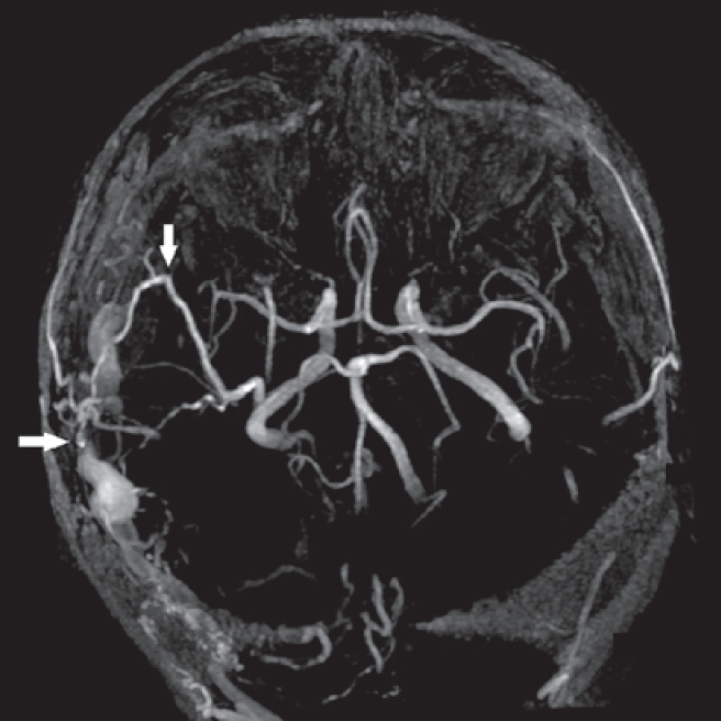
A 70-year-old man was admitted to our neurology department because of acute-onset left side hemiparesis. Neurological examination revealed anisocoria (right>left) with normal reaction to light, neck stiffness and bilateral Kernig sign, left side mouth asymmetry, paralysis of the left upper limb and severe paresis of the left lower limb with hypotonia and weak deep tendon reflexes, no pyramidal signs. Brain magnetic resonance angiography revealed an intracranial dural arteriovenous fistula [Figure - 1] and [Figure - 2]. The patient did not agree for endovascular treatment.
 |
| Figure 1: (a) T2-weighted fast spin-echo image showing hyperintense zone of venous congestion/ vasogenic oedema located in the right parietal lobe (black arrow) and widened right vein of Labbé and thalamostriate (white arrows). (b) Susceptibility- weighted imaging reveals zone of oedema (horizontal arrow), and subcortical microbleedings (vertical arrow) due to venous congestion/ hypertension. (c) T1-weighted image after contrast media administration shows enhanced widened, tortuous right vein of Labbé. (d) T1-weighted image after contrast, maximum intensity projection reveals widened, tortuous veins of right hemisphere: vein of Labbé, thalamostriate and parenchymal (arrows). |
 |
| Figure 2: 3D time of flight arterial phase magnetic resonance angiography, maximum intensity projection reconstruction. Widened right middle meningeal artery (vertical arrow) feeding dural arteriovenous fistula located in the right occipitotemporal region. The horizontal arrow indicates dural arteriovenous fistula and dilated, tortuous arterialized right vein of Labbe, and superficial veins of venous drainage. |
A massive angiogenic ipsilateral hemispheric cerebral oedema caused by an antidromic flow in cortical veins is a rare complication of dural arteriovenous fistula.[1]
| 1. | Dong Y, Cao W, Huang L, Li L, Zhang Y, Dong Q, etal. Ipsilateral intracranial edema associated with drainage patterns of dural arteriovenous fistula. J Stroke Cerebrovasc Dis 2014;23:1094-8. [Google Scholar] |
Fulltext Views
1,802
PDF downloads
1,706


