Translate this page into:
Wilson disease: Copper in the eye
Corresponding Author:
Pradeep Venkatesh
Dr Rajendra Prasad Centre for Ophthalmic Sciences, All India Institute of Medical Sciences, New Delhi
India
venkyprao@yahoo.com
| How to cite this article: Takkar B, Temkar S, Venkatesh P. Wilson disease: Copper in the eye. Natl Med J India 2018;31:122 |
An 8-year-old boy presented with a history of change in voice, difficulty in swallowing and abnormal posturing of hands and neck for 3 months. The parents also noticed that the boy was restless and irritable. Birth, developmental and family histories were unremarkable. On examination, dystonia of both hands and neck was noted. Motor examination showed rigidity of the left upper and lower limbs with exaggerated deep tendon reflexes. The boy did not cooperate during assessment of higher mental functions.
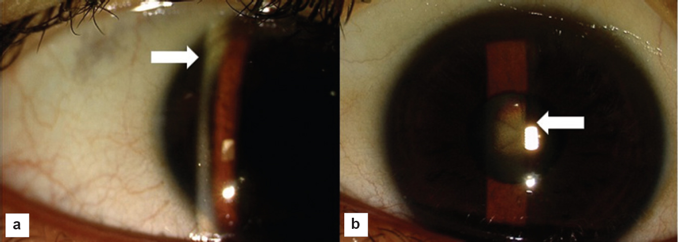
Ocular examination under magnification revealed greenish-brown-coloured ring at the level of the Descemet membrane in both peripheral corneas (arrow), suggestive of a Kayser-Fleischer ring [Figure - 1]a. The ring was distributed regularly all around the limbus. Further examination of the lenses showed ‘sunflower’ cataracts (arrow; [Figure - 1]b). Fundus examination was normal. Thus, a clinical diagnosis of Wilson disease was made. Investigations revealed elevated serum copper levels, low serum ceruloplasmin levels and grossly elevated urine copper levels confirming the diagnosis.
 |
| Figure 1: (a) Deposition of copper in the Descemet membrane of the cornea can be seen in the clinical photograph. Greenish-brown, ring-shaped deposition near the limbus is typical of a Kayser–Fleischer ring. (b) The arrow indicates a posterior subcapsular cataract. The cataract has a bright yellow sheen and is in the shape of a sunflower. |
The Kayser-Fleischer ring typically starts without symptoms at the vertical poles of the cornea due to deposition of copper in its deeper layers and then progresses circumferentially. Although early stages require a magnified examination, later stages may also be visible to the naked eye as a golden-brown ring.[1] The ring disappears as serum copper levels become normal with treatment.[1] The ‘sunflower’ cataract represents chalcosis of the lens, much like the one caused by intraocular copper foreign bodies. It may decrease with treatment though visually significant opacities require surgery.[2]
| 1. | Schrag A, Schott JM. Images in clinical medicine. Kayser-Fleischer rings in Wilson’s disease. N Engl J Med 2012;366:e18. [Google Scholar] |
| 2. | Yap EY, Buettner H. Spontaneous resolution of a sunflower cataract. Am J Ophthalmol 1992;113:723-5. [Google Scholar] |
Fulltext Views
2,210
PDF downloads
2,192


